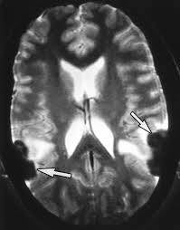
Sarcoidosis and Brain Fog: An Introduction
Brain fog describes problems with concentration, memory, word-finding or mental clarity. In sarcoidosis it may be related to fatigue, poor sleep, pain, medication effects, mood, inflammation or nervous-system involvement.
Keep a symptom diary and discuss persistent or worsening cognitive symptoms with your doctor. Treatable causes should be assessed before attributing every problem to sarcoidosis.
Translation of the original Croatian article
Source: original Croatian article.
Sarcoidosis, neuroinflammation and mild narrowing of the gyri and sulci of the left hemisphere: association with brain fog and options for a therapeutic approach
1. Introduction
We present the case of a 45-year-old man from Zagreb diagnosed with atypical, chronic sarcoidosis that developed over several years into a multisystem form of the disease. In this patient, sarcoidosis manifests not only through respiratory symptoms, but also through pronounced rheumatological, neurological and cardiac manifestations, which significantly impair quality of life and functional abilities.
The clinical course includes:
rheumatological manifestations: daily pain in the joints of the hands, ankles and hip, synovitis and tenosynovitis, morning stiffness, a need for walking aids;
neurological manifestations: small-fibre polyneuropathy, radiculopathy, autonomic dysfunction, chronic pain syndrome, mild narrowing of the gyri and sulci of the left hemisphere on MRI;
cardiovascular manifestations: episodes of hypoxaemia (saturation down to 80%), chest and jaw pain, elevated troponin (maximum value 34.8 ng/L), with normal coronary angiography and cardiac ultrasound, which raises suspicion of cardiac sarcoidosis or myocarditis; general condition: loss of more than 13 kg within a short period, reduced exercise tolerance and exhaustion after minimal physical activity.
This case clearly shows that sarcoidosis, although a rare disease, can take an atypical, chronic and progressive form, with multiple-organ involvement. The neurological manifestations are of particular importance, as these are the ones associated with the occurrence of cognitive disorders and the subjective feeling of so-called brain fog. The radiological finding of mild narrowing of the gyri and sulci of the left hemisphere is a subtle but clinically significant sign of chronic neuroinflammation and a potential basis for the development of lasting cognitive difficulties.
2. Pathophysiological basis: how sarcoidosis affects the brain
The pathogenesis of neurosarcoidosis is based on a T-lymphocyte-mediated immune response. There is activation of CD4+ T lymphocytes, accumulation of macrophages and formation of granulomas, secretion of proinflammatory cytokines such as IL-6, TNF-α and IFN-γ, and chronic activation of microglia in the CNS.
Such neuroinflammation leads to damage to nervous tissue, reduced synaptic plasticity and disruption of the brain’s functional networks. In the long term, this can manifest as cortical atrophy visible on MRI.
Mild narrowing of the gyri and sulci of the left hemisphere is precisely one of these radiological indicators: it means that thinning of the cortex has occurred as a result of a chronic process.
3. Radiological aspect: what “mild narrowing of the gyri and sulci” means
On brain MRI, the gyrus (fold) and sulcus (groove) show the structure of the cerebral cortex. Mild narrowing means that the folds are [Source-error note: the Croatian word “užljepljene” appears here and is not understandable in this context; its meaning remains unresolved and has not been reconstructed.] and the grooves are shallower, which is a sign of a reduction in the volume of cortical tissue.
Possible causes:
chronic neuroinflammation (sarcoidosis, multiple sclerosis, vasculitis), a long-term degenerative process, post-inflammatory changes following an earlier episode.
In sarcoidosis, this finding most often means that the inflammatory process in the brain has been long-lasting and has left a mark on the morphology of the cortex.
Why the left hemisphere? The left hemisphere dominates the processes of language, verbal memory and attention. Changes in this part of the brain can therefore explain the occurrence of symptoms such as slowed speech, difficulty concentrating and so-called brain fog.
4. Brain fog: definition and symptoms
Brain fog is not an official diagnosis, but a term used by patients themselves to describe their experience. It most commonly occurs in autoimmune and inflammatory diseases (sarcoidosis, multiple sclerosis, lupus, fibromyalgia).
Main symptoms:
-Difficulty concentrating and maintaining attention
-Forgetfulness (especially short-term memory)
-Slowed thinking and difficulty finding words
-Reduced ability to multitask
-Mental fatigue after simple tasks
-A feeling of “fog” in the head, as if thoughts are not clear
Unlike depression or dementia, brain fog is:
-fluctuating (stronger on some days, weaker on others),
-often associated with fatigue, illness or inflammation,
-reversible to some extent (it improves when the underlying disease is controlled or with mental exercises).
5. Mechanisms of brain fog in sarcoidosis
1. Inflammatory cytokines
– IL-6 and TNF-α cross the blood–brain barrier and affect neurotransmission, causing fatigue and cognitive difficulties.
2. Cortical atrophy
– mild narrowing of the gyri and sulci reflects a loss of cortical neurons, which manifests clinically as impaired attention and slowness.
3. Impaired hippocampal function
– the hippocampus is essential for memory and orientation. Neuroinflammation damages synaptic plasticity and leads to forgetfulness.
4. Autonomic dysfunction
– damage to the vagus nerve and the autonomic nervous system disrupts the regulation of blood pressure and cerebral perfusion, which worsens the feeling of “fog”.
6. Clinical evidence and literature
Studies have shown that patients with sarcoidosis have cognitive difficulties more often than the healthy population, even when there are no major structural lesions.
Fritz et al. (2016) – a meta-analysis showed that 30–50% of patients with neurosarcoidosis develop cognitive symptoms.
Hoitsma et al. (2004) – brain fog and neurocognitive disorders may be an early sign of neurosarcoidosis, often before major lesions develop.
Tana et al. (2022) – systemic neuroinflammation (including sarcoidosis) is associated with the feeling of brain fog through cytokine dysfunction.
7. Therapeutic implications
1. Corticosteroids – the first choice, but they lead to side effects in the long term.
2. Immunosuppressants (MTX, Arava) – often used, but not always effective.
3. Biological therapy (adalimumab, infliximab) – recommended for resistant forms, and increasing evidence supports its effectiveness in neurosarcoidosis.
4. Supportive measures – sleep hygiene, physical exercise adapted to the person’s abilities, cognitive rehabilitation.
8. Concentration exercises to reduce brain fog
Although the inflammatory process requires medical treatment, the affected person can use their own strategies to reduce the subjective difficulties of brain fog.
Mental exercises
-Mindfulness meditation – trains attention to focus on the present moment and reduces distractions.
-Cognitive games (memory cards, Sudoku, crosswords) – promote neuroplasticity and strengthen synaptic connections.
-The 5–10-minute concentration technique – choosing one activity and consciously focusing on it without interruption.
Organisational strategies
-Writing notes and using planners – reduces the load on working memory.
-Breaking tasks into smaller steps – reduces the feeling of being overwhelmed.
-A break ritual – short breaks every 30–40 minutes reduce mental fatigue.
Physical–cognitive synergy
-Breathing and light exercises – improve oxygenation of the brain.
-Short walks – stimulate circulation and concentration.
-Good sleep – regulates memory consolidation and reduces fatigue.
9. Long-term consequences and the importance of follow-up
If neuroinflammation is not treated, mild narrowing of the gyri and sulci can progress to more pronounced cortical atrophy. In the long term, this increases the risk of:
-permanent cognitive deficits,
-reduced capacity for work,
-social dysfunction and depression.
Regular MRI follow-up, monitoring of cognitive functions and timely initiation of biological therapy reduce the risk of progression.
10. Conclusion
Mild narrowing of the gyri and sulci of the left hemisphere in a patient with sarcoidosis is neither an incidental nor an insignificant finding. It is an indicator of chronic neuroinflammation and a possible initial stage of cortical atrophy. Clinically, it can manifest as brain fog, whose symptoms significantly impair quality of life.
A combination of medical treatment (biological medicines) and mental–cognitive exercises (mindfulness, cognitive games, organisational strategies) can alleviate subjective difficulties and preserve cognitive function.
References
1. Lower EE, Broderick JP, Brott TG, Baughman RP. Diagnosis and management of neurosarcoidosis. Arch Intern Med. 1997;157(16):1864–1868.
2. Fritz D, van de Beek D, Brouwer MC. Clinical features, treatment and outcome in neurosarcoidosis: systematic review and meta-analysis. BMC Neurol. 2016;16(1):220.
3. Ungprasert P, Matteson EL. Neurologic manifestations of sarcoidosis. Continuum (Minneap Minn). 2017;23(3):751–767.
4. Hoitsma E, Faber CG, Drent M, Sharma OP. Neurosarcoidosis: a clinical dilemma. Lancet Neurol. 2004;3(7):397–407.
5. Tana C, Donatiello I, Caputo A, et al. Brain fog in internal medicine: a comprehensive review on systematic disease-related causes and mechanisms. Intern
Emerg Med. 2022;17(1):1–17.
6. Judson MA. Neurosarcoidosis: clinical manifestations, diagnosis and treatment. Clin Chest Med. 2015;36(4):643–656.
Dino-Josip Ključarić, mag.theol.