WHAT ARE GRANULOMAS CAUSED BY SARCOIDOSIS?

Mechanism of granuloma formation
(Click to enlarge the image)
Description of granulomas
Granulomas are characteristic signs of symptomatic sarcoidosis. They are relatively small groups of inflamed cells with intense proliferative activity.
The original description characterises granulomatous lesions as unmistakably distinguishing sarcoidosis. On laboratory examination, it divides a granuloma into central and peripheral regions:
Central zone → rich in macrophages (at different stages of activation and differentiation) and Langerhans cells surrounded by CD4 lymphocytes (or T-helper cells).
The description also includes epithelioid cells (derived from macrophages) and giant cells with Schaumann bodies (cells with phosphatase).
Peripheral zone → rich in CD8 (suppressor) lymphocytes. It also contains antigen-presenting cells (macrophages) and plasma cells. The antigen binds as a key to the appropriate receptor.
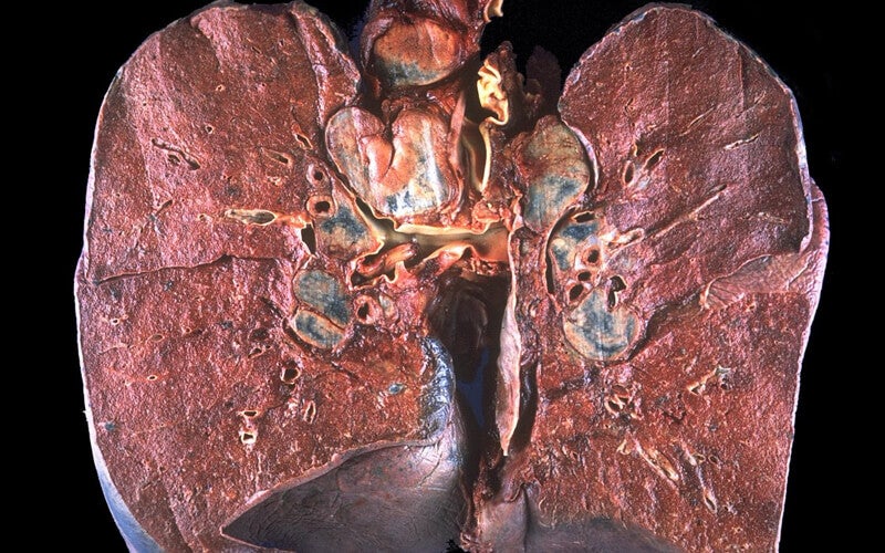
Although granulomas can occur in any anatomical location, they tend to originate in the lungs and then spread to areas such as the skin, central nervous system, eyes, heart, lymph nodes and other organs.
Kidneys, liver and spleen are also included among the sites listed in the source.
Granulomas may be completely reabsorbed, disappearing over a shorter or longer period of months or years without permanent damage, or they may create irreversible lesions. In the latter case, sarcoidosis follows a chronic course: the granulomas cannot be spontaneously reabsorbed and the affected tissue is damaged. The resulting damage is proportional to their spread and depends on the region involved. The source stresses that involvement of important organs such as the heart can be extremely dangerous and, without prompt intervention, can be fatal.
The source reports that sarcoidosis is fatal in only a small proportion of cases (5–10%).
The image shows granulomas and lesions in the lungs
(Click to enlarge the image)